Suikerrijke voeding veroorzaakt de ontwikkeling van alvleesklierkanker en verhoogt de kans op een dodelijke afloop. Tot die bevinding zijn onderzoekers van het Vlaams Instituut voor Biotechnologie (VIB) en de KU Leuven gekomen.
Een suikerrijk dieet verhoogt niet alleen de kans op het ontwikkelen van pancreaskanker. Het stimuleert ook de agressieve groei van tumoren. Tot die conclusie komen onderzoekers van VIB-KU Leuven en collega's van het Babraham Institute van de Britse universiteit Cambridge.
De resultaten zijn gepubliceerd in het tijdschriftCell Reports.
Alvleesklierkanker is een zeldzame maar dodelijke vorm van kanker door late detectie en een slecht begrip van risicofactoren. Bekende risicofactoren zijn obesitas, voeding en diabetes type 2, maar door de lage incidentie en de onderlinge samenhang is hun individuele bijdrage moeilijk in te schatten.
De onderzoekers voltooiden een uitgebreid project met experimenteel werk bij muizen en menselijke gegevens. Bij de dieren werd de progressie van pancreaskanker beïnvloed door voedingssuiker, met een snellere tumorgroei en verhoogde dodelijkheid.
Het effect bij mensen blijkt hetzelfde. Bij 500 deelnemers aan de studie onderzochten de onderzoekers de interactie tussen genen en voeding en ontdekten dat hoge niveaus van voedingssuiker het risico op alvleesklierkanker verhoogden. 'We hebben jarenlang gekeken naar verschillende voedings- en genetische veranderingen en niets komt in de buurt van de nadelige gevolgen van een dieet met veel suiker', zegt James Dooley, senior wetenschapper bij het Babraham Institute.
Plantaardige voeding met een equivalent van een avocado per dag vermindert het risico op pancreaskanker met tien procent.
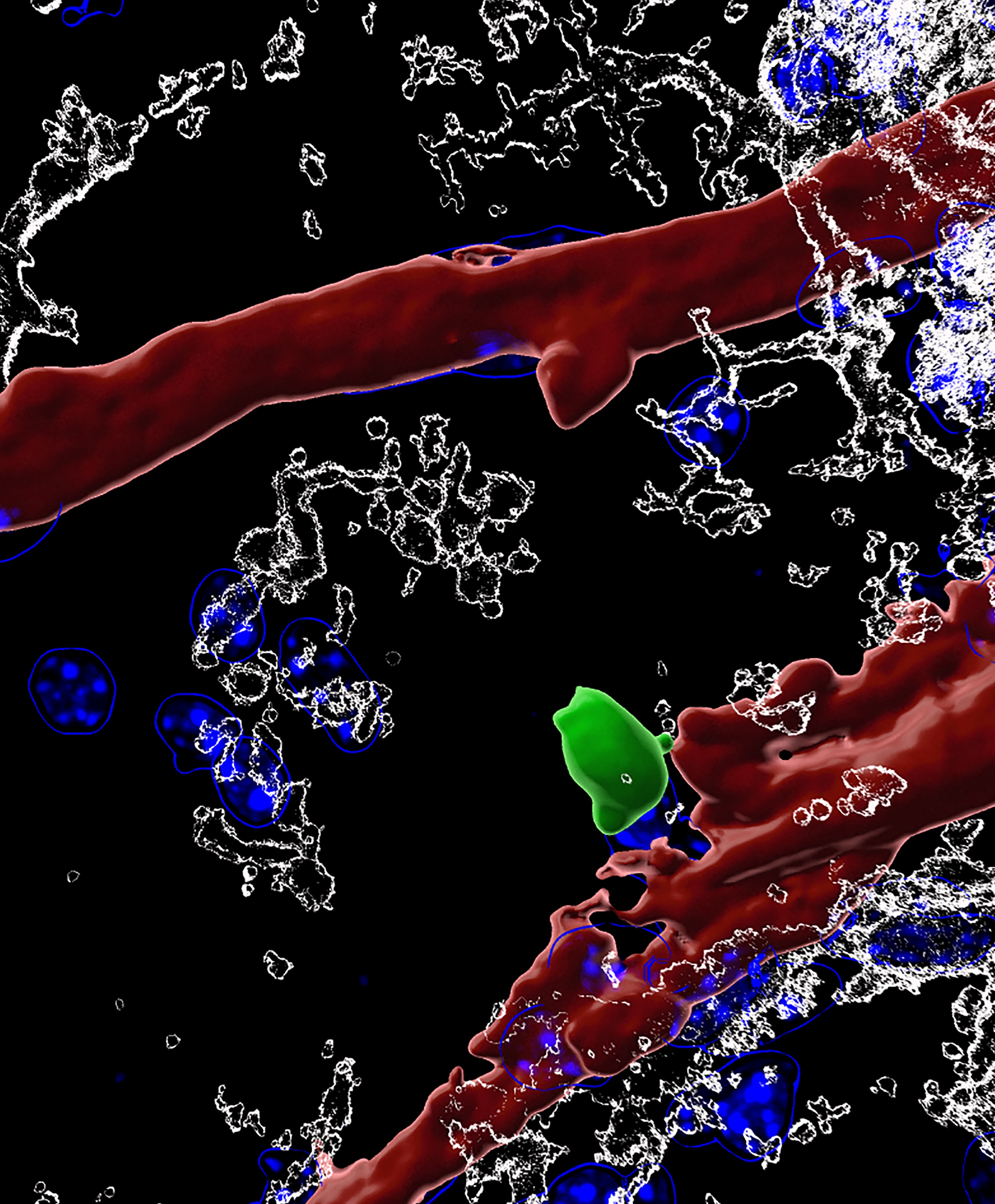
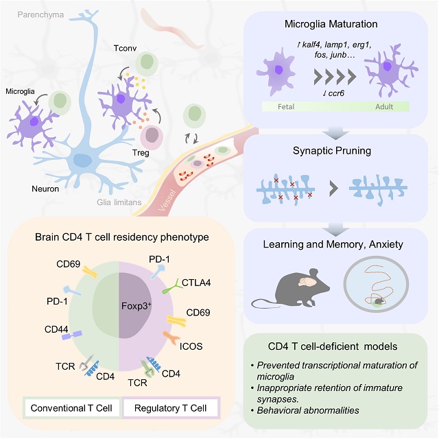
Whether white blood cells can be found in the brain has been controversial, and what they might be doing used to be complete mystery. In a seminal study published in Cell, an international team of scientists led by Prof. Adrian Liston (VIB-KU Leuven, Belgium & Babraham Institute, UK) describe a population of specialized brain-resident immune cells discovered in the mouse and human brain, and show that the presence of white blood cells is essential for normal brain development in mice.
Like a highly fortified headquarters, our brain enjoys special protection from what is circulating in the rest of our body through the blood-brain barrier. This highly selective border makes sure that passage from the blood to the brain is tightly regulated.
The blood-brain barrier also separates the brain from our body’s immune system, which is why it has its own resident immune cells, called microglia, which trigger inflammation and tissue repair. Microglia arrive in the brain during embryonic development, and later on, the population becomes self-renewing.
Yet, white blood cells—which are part of our immune system—have been found to play a role in different brain diseases, including multiple sclerosis, Alzheimer’s and Parkinson’s disease or stroke. Whether or not white blood cells can be found in healthy brains as well, and what they might be doing there, has been subject of intense debate. An interdisciplinary team of scientists led by Prof. Adrian Liston (VIB-KU Leuven, Babraham Institute) set out to find the answers.
White blood cells in the brain
"A misconception about white blood cells comes from their name,” explains Dr. Oliver Burton (Babraham Institute). “These 'immune cells' are not just present in the blood. They are constantly circulating around our body and enter all of our organs, including—as it turns out—the brain. We are only just starting to discover what white blood cells do when they leave the blood. This research indicates that they act as a go-between, transferring information from the rest of the body to the brain environment"
The team quantified and characterized a small but distinct population of brain-resident T helper cells present in mouse and human brain tissue. T cells are a specific type of white blood cells specialized for scanning cell surfaces for evidence of infection and triggering an appropriate immune response. New technologies allowed the researchers to study the cells in great detail, including the processes by which circulating T cells entered the brain and began to develop the features of brain-resident T cells.
Dr. Carlos Roca (Babraham Institute): “Science is becoming increasingly multidisciplinary. Here, we didn't just bring in expertise from immunology, neuroscience and microbiology, but also from computer science and applied mathematics. New approaches for data analysis allow us to reach a much deeper level of understanding of the biology of the white blood cells we found in the brain.”
An evolutionary role
When T helper cells are absent from the brain, the scientists found that the resident immune cells – microglia – in the mouse brain remained suspended between a fetal and adult developmental state. Observationally, mice lacking brain T cells showed multiple changes in their behavior. The analysis points to an important role for brain-resident T cells in brain development. If T cells participate in normal brain development in mice, could the same be true in humans?
“In mice, the wave of entry of immune cells at birth triggers a switch in brain development,” says Liston. “Humans have a much longer gestation than mice though, and we don't know about the timing of immune cell entry into the brain. Does this occur before birth? Is it delayed until after birth? Did a change in timing of entry contribute to the evolution of enhanced cognitive capacity in humans?”
The findings open up a whole new range of questions about how the brain and our immune system interact. "It has been really exciting to work on this project. We are learning so much about how our immune system can alter our brain, and how our brain modifies our immune system. The two are far more interconnected than we previously thought," says Dr. Emanuela Pasciuto (VIB-KU Leuven).
The study also brings in a connection with the gut microbiome, says Liston: “There are now multiple links between the bacteria in our gut and different neurological conditions, but without any convincing explanations for what connects them. We show that white blood cells are modified by gut bacteria, and then take that information with them into the brain. This could be the route by which our gut microbiome influences the brain.”
Taken together, the results contribute towards the increasing recognition of the role of immune cells in the brain and shed new light on its involvement in a range of neurological diseases.
Witte bloedcellen maken deel uit van ons immuunsysteem dat ons tegen ziektes beschermt. Of ze ook in de hersenen terug te vinden zijn, en wat ze daar dan zouden doen bleef tot nog toe een raadsel. Een internationaal team van wetenschappers onder leiding van professor Adrian Liston (VIB-KU Leuven, België en Babraham Institute, VK) toont nu aan dat witte bloedcellen wel degelijk voorkomen in de hersenen van zowel muizen als mensen, en dat hun aanwezigheid essentieel is voor normale hersenontwikkeling. De resultaten verschijnen deze week in het prestigieuze vakblad Cell.
Onze hersenen worden als een versterkte burcht ommuurd door de bloed-hersenbarrière. Die moet vermijden dat stoffen die in onze bloedbaan circuleren zomaar in onze hersenen terechtkomen. Via de bloed-hersenbarrière worden onder strikte controle enkel welbepaalde stoffen uitgewisseld.
De bloed-hersenbarrière schermt de hersenen ook af van ons immuunsysteem, dat de rest van ons lichaam patrouilleert om bijvoorbeeld bacteriële of virale indringers op te sporen en uit te schakelen. Precies daarom heeft het brein z’n eigen immuuncellen: microglia.
Toch blijken witte bloedcellen ook een rol te spelen bij verschillende hersenaandoeningen. Denk maar aan MS, alzheimer, parkinson of een beroerte. Hierbij gaat het wel telkens om ziek of ‘beschadigd’ hersenweefsel, waar mogelijk ook de bloed-hersenbarrière is aangetast. De vraag bleef dus of – en waarom – witte bloedcellen nu werkelijk aanwezig zijn in hersenen die normaal en gezond zijn.
Witte bloedcellen in de hersenen
Een interdisciplinair team van wetenschappers onder leiding van prof. Adrian Liston (VIB-KU Leuven, Babraham Institute) heeft nu een kleine maar belangrijke groep van T-helpercellen ontdekt in hersenweefsel afkomstig van muizen en van mensen. T-helpercellen zijn een specifiek type witte bloedcellen, gespecialiseerd in het scannen van celoppervlakken op aanwijzingen van infectie en in het op gang trekken van een aangepaste immuunreactie. Aan de hand van de laatste technologie konden de wetenschappers de T-helpercellen tot in detail bestuderen, inclusief hoe en wanneer ze in de hersenen terecht komen.
Dr. Emanuela Pasciuto (VIB-KU Leuven), postdoctoraal onderzoeker in het team van Liston benadrukt het belang van interdisciplinair onderzoek: “Om de rol van witte bloedcellen in het brein in kaart te brengen hebben we niet alleen expertise van immunologie, neurowetenschappen en microbiologie bij elkaar gebracht, maar ook van informatica en toegepaste wiskunde. Nieuwe benaderingen voor data-analyse stellen ons in staat om een veel dieper begrip te krijgen van de biologie van de witte bloedcellen die we in de hersenen hebben gevonden.”
Een evolutionaire rol
De onderzoekers stelden vast dat in muizenhersenen zonder T-helpercellen de ontwikkeling van de typische immuuncellen van het brein (de microglia) bleef hangen ergens tussen een foetale en volwassen ontwikkelingsstatus. De muizen zonder T-helpercellen in de hersenen vertoonden bovendien verschillende gedragsafwijkingen, wat wijst op een belangrijke rol voor de T-helpercellen bij de normale hersenontwikkeling. En als dat geldt voor muizen, zou hetzelfde dan ook waar zijn voor mensen?
“We zien dat de toestroom van immuuncellen in de hersenen bij de geboorte van muizen leidt tot een omslag in het ontwikkelingsproces,” zegt Liston. “Maar de zwangerschap bij mensen is veel langer dan bij muizen, en we weten niet wanneer de immuuncellen dan toekomen in het menselijk brein. Gebeurt het nog vóór de geboorte? Is het uitgesteld tot na de geboorte? Kan een verandering in de timing bijgedragen hebben aan de evolutie van de uitzonderlijke hersencapaciteit van mensen?”
De bevindingen openen een heel nieuw gamma aan vragen over de wisselwerking tussen ons brein en ons immuunsysteem. “We leren nog elke dag bij over hoe ons immuunsysteem ons brein kan beïnvloeden en vice versa. De twee zijn veel meer met elkaar verbonden dan we eerder dachten,” zegt Pasciuto.
Darmen en hersenen
De studie legt ook nieuwe verbanden tussen ons brein en onze darmflora, aldus Liston: “Heel wat neurologische aandoeningen worden in verband gebracht met bacteriën in onze darmen, maar zonder overtuigende verklaringen voor die connectie. Onze resultaten laten zien dat darmbacteriën witte bloedcellen kunnen beïnvloeden, die deze ‘informatie’ vervolgens mee nemen naar de hersenen. Dit zou de manier kunnen zijn waarop onze darmflora onze hersenen beïnvloeden. ”
De nieuwe resultaten dragen enorm bij tot de groeiende kennis over de rol van immuuncellen in de hersenen, zowel tijdens de normale ontwikkeling als bij verschillende ziekteprocessen.
Research using mice and human data shows how diet affects disease risk
Eating a diet high in sugar increases the likelihood of developing pancreatic cancer in some individuals and also drives the aggressive growth of tumours, a study by researchers from the Babraham Institute, Cambridge, UK, and VIB-KU Leuven, Belgium, has found.
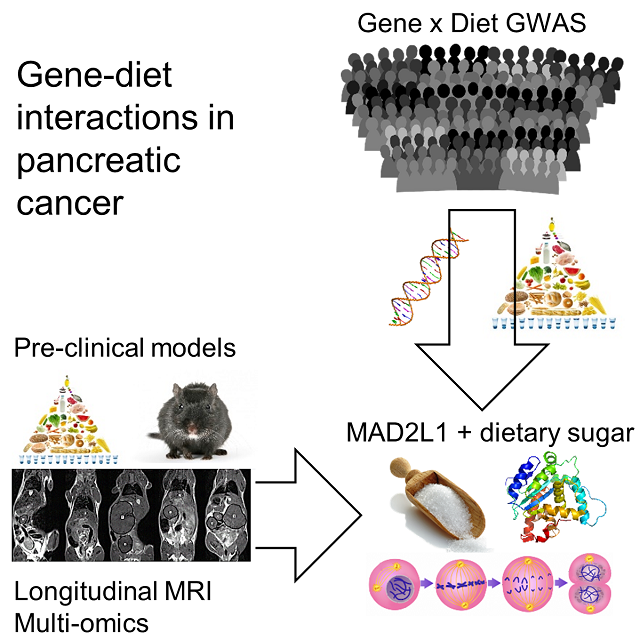
The researchers completed a comprehensive project using experimental work in mice and human data, including from pancreatic cancer patients, to understand the influence of different dietary components on the development and progression of pancreatic cancer. The research is published today in the journal Cell Reports.
Pancreatic cancer is a rare but fatal form of cancer, due to late detection and a poor understanding of the risk factors. Known risk factors include obesity, diet and type 2 diabetes, however the low incidence rate and interconnection of these factors mean that it is difficult to tease apart their individual contribution.
The researchers first studied the effects of obesity, diet and diabetes on pancreatic cancer development, growth and lethality in mice. In parallel they analysed the effect of diet using data from the European Prospective Investigation into Cancer and Nutrition (EPIC) study, which followed over half a million Europeans for 20 years.
The researchers found that obesity, diet and diabetes had profound and differing impacts on cancer incidence and growth. Using mice, the results indicated that obesity, dietary animal fats and dietary sugar were independent drivers of different facets of pancreatic cancer progression. In particular the results shed light on how pancreatic cancer might be affected by dietary sugar, with more rapid tumour growth and escalated lethality.
The effect of dietary sugars on pancreatic cancer development was preserved between mice and humans. In 500 study participants with pancreatic cancer, the researchers explored the interaction between genes and diet and found that high levels of dietary sugar increased pancreatic cancer risk in individuals with a certain genetic variation (found in 6% of the population).
Dr James DooleyDr James Dooley, senior staff scientist in the Immunology programme at the Babraham Institute, said: "Our study raises concern about the remarkable toxicity of sugar in our diet. We spent years looking at different dietary and genetic changes, and nothing has anywhere near the detrimental impact of a high sugar diet. Our findings suggest that it drives pancreatic cancer onset and makes it a more aggressive and lethal tumour."
Analysis of the human-derived data from the large EPIC study suggested that dietary plant fats reduced the risk of pancreatic cancer, estimating a 10% decrease in risk when eating the equivalent of an avocado a day.
Professor Adrian Liston, senior group leader at the Babraham Institute, said: "This study shows the power of combining animal research with the study of patients. We were able to use epidemiology and large patient-based resources to find a link in humans, and then go back to an animal model to formally test the direction of causality. Finding the same gene-diet link in both mice and humans makes us confident that diet is modifying disease risk, and gives us the tools to test preventative and therapeutic interventions."
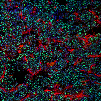
Proliferating pancreatic cancer cells in mice fed high sugar dietsWhile the impact of nutrition on cancer development continues to be explored and debated, this study provides a vital foundation for further studies and important indications to explore. Armed with this knowledge, clinicians could be supported to identify individuals at increased risk for screening and individuals with a high-risk genetic background could take pro-active dietary changes to reduce their risk of pancreatic cancer.
Ali Stunt FRSA, Founder and Chief Executive of Pancreatic Cancer Action, concurs: "Here at Pancreatic Cancer Action, we strongly advocate for high quality research into this neglected cancer. Dr Liston and his team have made a major contribution into understanding how diet and genetics changes the risk for pancreatic cancer. Healthy lifestyle choices, such as avoiding sugar-sweetened beverages and choosing a diet rich in vegetables, can reduce your risk of pancreatic cancer, and may be especially important in families with a history of the disease."
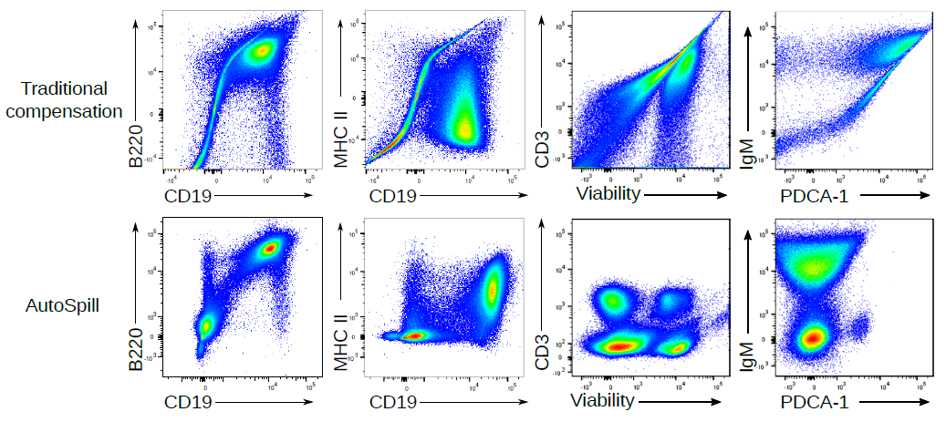
I am really thrilled to release AutoSpill onto BioRxiv. AutoSpill is a novel method for applying compensation to flow cytometry data, which reduces the error by ~100,000-fold. It is thanks to AutoSpill that we can push machines to their max colours, and actually get good quality 40+ parameter flow cytometry data. AutoSpill is a beautiful example of what maths can add to #immunology, led by the talented Dr Carlos Roca.
So how does AutoSpill work? If you just want to compensate your data, simply upload your single colour controls to https://autospill.vib.be and then copy the spillover matrix to your flow cytometry program of choice. Dr Carly Whyte made this easy two minute tutorial:
If you program your flow cytometry analysis in R, we have also released the AutoSpill full code, so you can add this to your bioinformatics pipeline.
Here are a few examples of the error reduction you can get with AutoSpill:
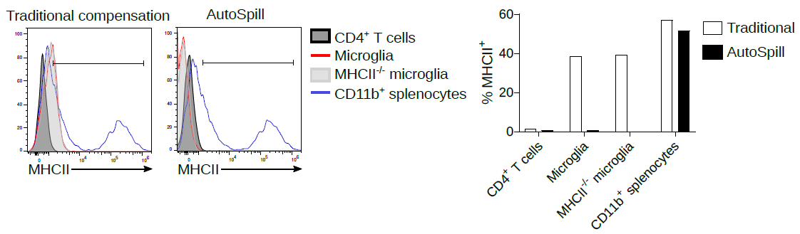
In high dimensional flow cytometry traditional compensation errors create artefacts. AutoSpill creates a perfect spillover matrix. What does a "perfect" spillover matrix mean? An error reduction of 100,000-fold on average, to the point where error is practically zero. (if you are using the script, you can reduce the error further - we stop the improvement at this point because it is functionally perfect). This means the large over/under-compensation effects can be completely removed from your data. If you want to run 28 colour flow cytometry on a 28 colour machine, you can spend hours-upon-hours compensating your data by hand, or 2 minutes with AutoSpill. AutoSpill is designed to run through the same operations that a skilled flow user does, just faster. But always remember - the compensation can still only be as good as the quality of the single colour controls!
How does AutoSpill work? It is a huge surprise to me, but with the enormous effort over decades to add extra lasers and new flurophores onto machines, the mathematics behind compensation hasn't been updated since 1993, where it was designed for 3 colour flow on computers with 100,000-fold less capacity. For decades we've been building more-and-more expensive machines, and haven't updated the basic maths that the machines run on!
Traditional compensation defines a positive and a negative population, finds the slope and uses that as the spillover matrix. It still works okay in most cases, it is just that the small errors start piling up when you are making 250+ calculations on a high parameter dataset.
AutoSpill is actually fairly simple at heart:
Draw an automatic live cell gate
Use linear regression to take into account every cell, not just the two data points of average positive and average negative
Calculate the error left, using the sum of errors in every compensation pair
Use the residual error and return to step 2
Repeat the tweaking of the matrix until error is gone
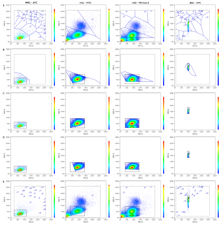
The underlying mathematics is tougher though (never thought I'd write a paper discussing "the linearity of the quantum mechanical nature of photons"!), because AutoSpill was built for actual flow cytometry users, not as a computational exercise. Carlos built the original pipeline, and then we extensively beta-tested it on 1000+ datasets over 20 months. Something as simple as a live gate becomes complex when you want it to robustly work on any dataset, cells or beads, collected on any machine. I'll spare you the details, but two stage tesselation and a 33% density estimation using a convex hull does the trick, successfully spotting the cells or beads even with heavy debris:
There are many advantages to using linear regression to calculate compensation. Why through out the data from 40,000 cells and instead turn it into two points, the way traditional compensation does? If you use linear regression you can use all of the data, which means AutoSpill works even if you have mostly negative or mostly positive data, just a shoulder of positive events or even a smear. So you can use the real antibodies on real cells to calculate your single colour compensations, rather than using beads or anti-CD4 in every channel.
As an added advantage to this approach, AutoSpill can remove most of the autofluroscence from your flow cytometry sample. For people working on cancer or myeloid cells this can be a complete game-changer. It turns out that while cells have different amounts of autofluorescence, the spectrum of that autofluirescence is fairly constant. You can collect empty data in the worst autofluorescent channel. The single in this channel can be used to calculate the autofluorescent spectrum, which can then be calculated on a per-cell basis and used to compensate it out of every other channel.
Here are two examples:
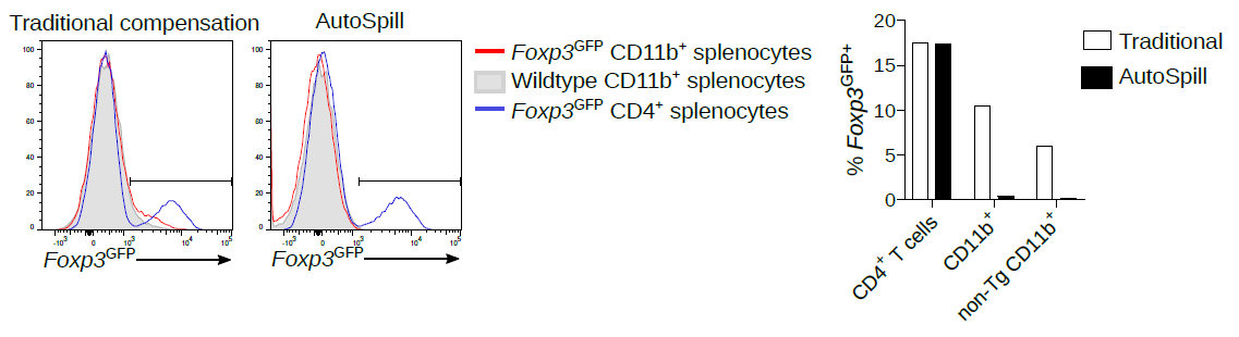
1. Back when I was a post-doc, there were many published reports of Foxp3 expression in macrophages, epithelium, cancer, etc. All autofluorescent artefacts that wasted years of research. AutoSpill removes this autofluorescence specifically from the macrophages:
2. Now we work on microglia, and it is still argued in the literature as to whether they express low levels of MHCII at homeostasis. Using AutoSpill to remove the autofluorescent signal it is quite simple - no they don't.
Of course, AutoSpill is a tool to get an optimal solution for good data. It can't turn bad data into good data. You should always work with your Core Facility staff to optimise the machines and run high quality single colour controls.
If you like AutoSpill, and you come from a math, computer science or data science background, why not come and join the lab? We have a position open for a data science post-doc or senior scientist for another two weeks.
This is a strange time for any workplace. People suddenly working from home, large changes in job duties, some people left without much to do while others are expected to manage whole new realms of bureaucracy while also continuing their full-time job. For us, as an immunology lab, this pandemic has an added dimension of peculiarity: our work is directly relevant to the ongoing situation.
Looking back on how we dealt with the outbreak, we were ahead of the curve. We put in place strict social distancing and work-from-home measures well before our institutes / government did (and, I would argue as an immunologist, our lab rules were more science-based than those later imposed on us). We also started a public education program on COVID-19, with an interactive Virus Outbreak simulator, an illustrated series translating scientific articles into lay language and even released a kid's book explaining Coronavirus (with special thanks to lab members Dr Teresa Prezzemolo, Julika Neumann and Dr Mathijs Willemsen for translating this into different languages).
We also had lab members head back to the clinic to help with the capacity issues created by COVID-19. Dr Frederik Staels and Dr Erika Van Nieuwenhove suspended research to increase their clinical duties, and Dr Stephanie Humblet-Baron and Dr Mathijs Willemsen were on-call in case the system was overwhelmed.
Silke Janssen, processing patient blood
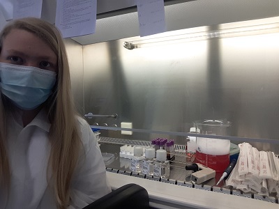
Our lab never completely shut-down though - we had important work that needed to be done. I'd like to call out Dr Susan Schlenner, Dr James Dooley and Dr Lubna Kouser who led the unglamorous but key administration on securing the safety of team members who had to be in the lab. Our Leuven lab was central to the processing of clinical COVID-19 samples. We usually think of clinical trials being run by MDs, but the work does not end after the blood is collected. I really want to call out the key contributions of Silke Janssens and Dr Teresa Prezzemolo. Without them, coming in all day, every day to process blood samples, clinical research of COVID-19 would have been crippled.
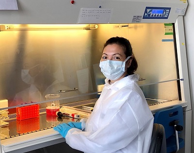
Dr Teresa Prezzemolo in the L2 labOur team, lead by Dr Stephanie Humblet-Baron, also analysed the samples prepared. We performed an ultra-high parameter analysis (far beyond state-of-the-art hospital diagnostics) of the T cell phenotype of COVID-19 patients: months of work from Dr Teresa Prezzemolo, Silke Janssens, Julika Neumann and Dr Mathijs Willemsen. Data analysis by Julika Neumann, Dr Carlos Roca, Dr Oliver Burton and Dr Stephanie Humblet-Baron identified a novel link between IL-10-producing Tregs and COVID-19 severity. We are now following this up to see if the link is useful as a biomarker or even is mechanistic in disease program. We have made our data an open resource (link), allowing other groups around the work to analyse our work. We are continuing to follow these patients and will soon have more and more information about why some patients remain completely healthy and others develop severe, even fatal, disease.
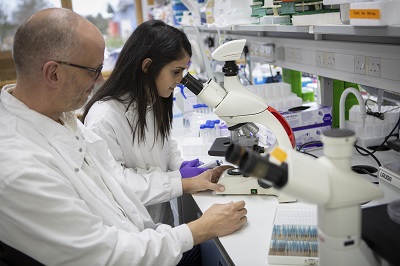
Dr Dooley and Dr Kouser (pre-COVID-19)We are not just clinical immunologists - we are also basic research immunologists. Mysterious virus triggering immune-mediated destruction of the tissue? We can deal with that. The whole lab contributed to the design of a new potential therapeutic, but I would especially like to call out the contributions of Dr James Dooley, Dr Oliver Burton, Dr Lubna Kouser and Fran Naranjo. Manufacturing is now complete and we are moving to pre-clinical testing. Hopefully we have a vaccine for SARS2 before our treatment is complete, but it is designed to deal with an unknown SARS3 equally well.
Suffice it to say, we have been as busy as we've ever been, and we will likely remain just as busy well after COVID-19 stops making the headlines. Which brings me to my final plea. Don't forget about scientific research. Unsung heroes during the pandemic, our staff are putting in an enormous effort. And yet we face an incredibly uncertain funding situation. Universities and research institutes have taken an enormous financial blow with this pandemic, and unless governments step in with a large financial rescue package, those scientific research staff who got us through the pandemic are going to be laid off in huge numbers. Even if you don't care about the moral imperative of looking after the people who stepped up when we needed them, there will be a SARS3 or novel flu pandemic in the future. We need to secure the research infrastructure to combat them right now. Science is not a factory that can be switched on and off at will - we need to maintain research excellence, scientific equipment and most of all key staff contracts over the long-term.
There is no use of mice except under the mouse user principles
There is no use of human samples or data without signing the patient data agreement
Racism, sexism and homophobia are not acceptable
Scientific fraud or plagerism are not acceptable
The laboratory is a shared facility with shared space and shared responsibility. As such
I have read and agree to follow the laboratory protocols
I will perform my lab duties as described in the “lab protocols” well and with regularity
When I go on holidays I will prearrange for my lab duties to be fulfilled by someone else
If I know in advance that performing my duties on schedule is not possible, I will consult with the lab manager on a case-by-case basis
Research laboratories can be a place of great stress, making inter-personal relationships intense. When dealing with other members of the laboratory:
I will try to treat every person with professional respect
I will try to be considerate of other’s feelings
I will try to be respectful of other’s working conditions (eg, noise, tidiness)
I understand that I work in a multicultural workplace, and that different people can have different cultural assumptions, reactions and methods of response than I do myself
I will try to assume the best rather than the worst from others’ actions
I will try to see the situation from the perspective of the other in a dispute
I understand that tensions will rise and that we will not always be perfect
When I err, I will apologise
When I am apologised to, I will try to forgive
I will try not to hold a grudge
I understand that public displays of affection or anger are not appropriate in the laboratory
When disputes arise, I understand there is a responsibility to keep the impact on others minimal, as well as a responsibility to resolve the problem
I understand that this code of conduct is an ambition for myself to achieve, rather than a standard against which I should judge others
Job opportunity: we need a junior lab technician at the University of Leuven to be trained for PBMC isolation and flow cytometry analysis, to place a key role in clinical trials. We are after someone who is willing to listen and takes their work seriously. If you already know flow cytometry, great, if not, we will train you. Apply here, and take on a job that matters.
Great opportunity available for a scientist to work on an exciting diabetes project in the Leuven lab. Pure cell biology / endocrinology, so we welcome applications from beyond immunology! We have a research technician and post-doc position available, depending on the experience / background of the applicant. If you are interested, please apply!


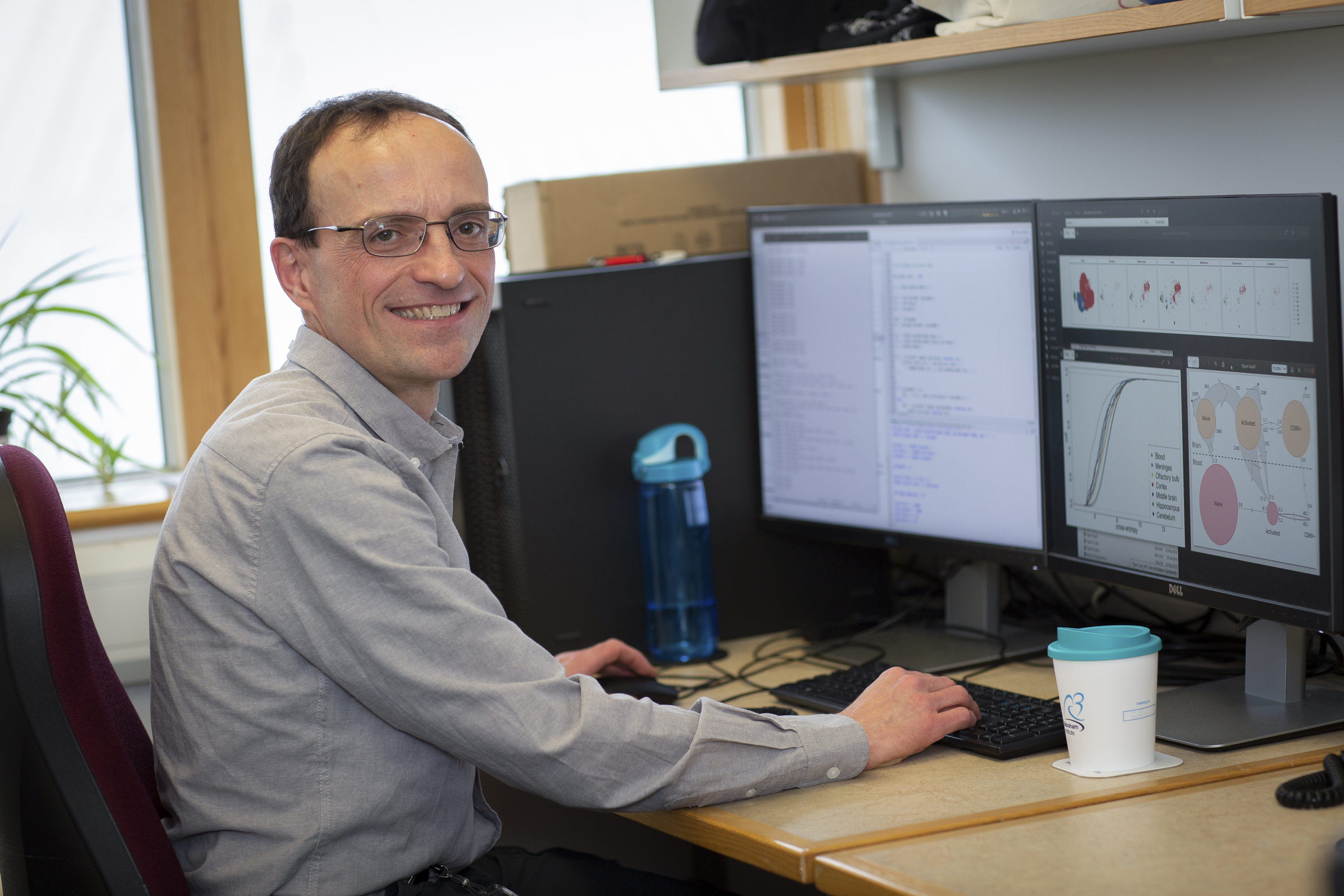
 Adrian Liston