Navigation
Public engagement

Becoming a Scientist

Read online for free

Print your own copy

Virus Fighter

Build a virus or fight a pandemic!

Play online

Maya's Marvellous Medicine

Read online for free

Print your own copy

Battle Robots of the Blood

Read online for free

Print your own copy

Just for Kids! All about Coronavirus

Read online for free

Print your own copy

Archive
LabListon on Twitter

Entries in neuroscience (21)

Tuesday
May092023

The key to healthy aging brains

As people age, they often experience problems with their memory and cognitive abilities. This happens in part because their brains become mildly inflamed. But there may be a solution: a small group of special T cells, called regulatory T cells, could help reduce this inflammation in aging brains. Administering a protein called interleukin-2 (IL2) can help these special T cells grow and prevent inflammation. Now, researchers at VIB, KU Leuven, Babraham Institute, and i3S have tested this approach in mice and found that it can prevent neurological decline. Their findings, published in EMBO Molecular Medicine suggest that targeting the immune system might keep people’s brains healthy as they age.

Emanuela Pasciuto, co-first author of the study: “Our goal was to see whether we could slow down the aging process of the brain by changing its immune system through the delivery of IL2. We know that inflammation plays a significant role in various aging processes, and IL2 could help us tilt the balance back in our favor.”

Inflammation in the aging brain

Aging is a degenerative process that affects the whole body, including the brain. As we age, our brains may experience cognitive decline, affecting our memory and ability to think clearly. Increasing evidence suggests that inflammation in the brain, called “inflammaging,” can worsen this decline. Inflammaging is caused by immune cells entering the brain as we age. This inflammation can activate microglia, the resident immune cells in the brain, and induce neuroinflammation, leading to cognitive decline and dementia.

However, researchers have found a way to reduce inflammation in the brain by targeting a small group of special immune cells in the brain called regulatory T cells. Previously, the team of Adrian Liston (VIB-KU Leuven, Babraham Institute) and Matthew Holt (VIB-KU Leuven, i3S Porto) showed that administering a protein called Interleukin-2 (IL2), which helps regulate the immune response, increased the number of regulatory T cells in the brain. This treatment has been successful in mouse models of traumatic brain injury and neuroinflammation.

Now, the researchers want to see if delivering IL2 directly to the brain can help reduce age-induced inflammation and cognitive decline.

Gene therapy improves brain aging

In their latest study, the team discovered that delivering IL2 to the brain improved brain function in aging mice. The research showed that the treatment restored cognitive performance in spatial memory tests, allowing older mice to form new memories almost as well as young mice. The mice given IL2 treatment were better at remembering visual cues than those that did not receive the treatment. Additionally, some of the changes in cellular aging in the brain were reversed, especially among several types of glial cells, which are critical to support overall brain function and health.

Pierre Lemaitre, co-first author of the study: “Our approach was to harness the body’s own system to regulate inflammation and to boost it precisely where it was needed.”

IL2 was delivered to the brain using a gene therapy vector, which is a tool that provides genetic material to specific cells. The additional dose of IL2 allowed the regulatory T cells to survive and create an anti-inflammatory environment. Matthew Holt and Lidia Yshii, co-senior authors of the study: “This reinforces our belief that viral vector-based systems are the way forward for the delivery of therapeutics to combat chronic neurodegenerative diseases and preventing cognitive decline in aging populations.”

Adrian Liston, senior author of the study: “The most important part of this study is the high potential for translation into patients. Inflammation is a process that is conserved in both mice and humans, and regulatory T cells can respond to IL2 in both species. However, there are still regulatory hurdles to clear, and it’s crucial to ensure safety before testing it in patients. Nonetheless, we see a clear path to conducting clinical trials.”

The laboratory is working through spin-off company Aila Biotech to drive entry of this therapeutic into clinical trials. Read the full study here

Monday
Jun202022

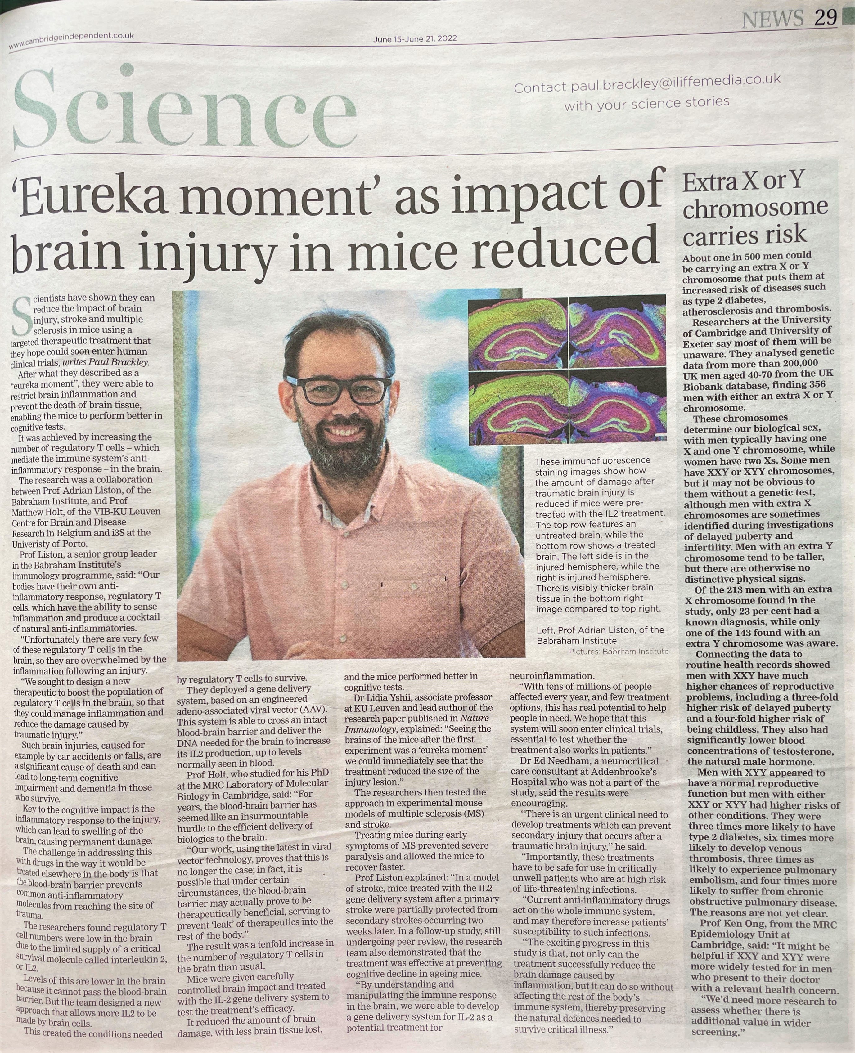
'Eureka moment' as impact of brain injury in mice reduced

Wednesday
Jun012022

Drugs, Vaccines and a Hopeful Future: Exploring Advances in Multiple Sclerosis Research

I was interested by Ruairi Mackenzie from Technology Networks for World Multiple Sclerosis Day. Here is the resulting article:


World Multiple Sclerosis (MS) Day recognizes the millions of people worldwide who are affected by this neuroimmunological disease. The campaign site for World MS Day 2022 strikes an optimistic chord, seeing the date as a chance to “celebrate global solidarity and hope for the future”. This year, there is more reason to buy into that optimism that ever before.

 Recognizing the immune basis of MS

Adrian Liston, a group leader at the Babraham Institute, based near Cambridge, UK, is well-placed to explain that sunny outlook. He first studied MS as part of an undergraduate project.  Over the two decades since, Liston has continued to research in the MS field, watching science’s understanding of the disease deepen.

“I think the most profound change has been the recognition that MS is an immune-mediated disease,” says Liston. The symptoms of MS are neurological – patients experience a range of sensory and motor conditions, including fatigue, numbness, spasms and weakness and loss of control over muscle movement or function. While MS has long been thought to include an immune component – Jean Martin Charcot, the French doctor who first described the disease, noted the presence of immune cells in patients’ spinal cords – it was only  recently that the immune-mediation of the disease was fully understood. It’s now recognized that the neurological symptoms are a consequence of immune cells infiltrating the brain and attacking the myelin sheath coating that permits normal nerve cell conduction.

Part of that understanding came from the galloping pace of genetics research. The neurological symptoms of MS are in contrast with its genetic signature, explains Liston. From a geneticist’s point of view, “MS looks a lot like Type 1 diabetes, or any of these other autoimmune diseases, and the same genes are controlling it,” he says.

These genetic insights were added to a rapidly growing body of preclinical research. Experimental autoimmune encephalomyelitis (EAE) is an inflammatory autoimmune disease seen in animals that mimics the disease course of MS. This model proved essential for the development of drugs for MS. Liston explains that these drugs came in two waves. The first was heralded by the approval of interferon beta-1b in 1993, the first drug capable of altering the course of MS. The interferons, which reduce the number of immune cells crossing the blood-brain barrier, showed success at improving MS symptoms. This provided direct evidence that adopting an immune-targeting approach could help patients.

A new generation of drugs

For some, these early drugs have proved enduringly beneficial and remain their main course of treatment years later. MS drug development, however, continued to refine the targeting of the immune system. “What we really we have now is the second wave of drug generation, where we have much more sophisticated immune-modulating molecules, and we can really target very specific pathways of cells that are causing the damage,” says Liston. These new drugs (there are 23 FDA-approved mediations for MS as of 2022) work better and for longer.

Liston says that an MS patient today can expect fewer symptoms and far longer periods of good health than in the past. 85% of MS patients have a relapsing-remitting form of the disease, which sees symptoms wax and wane. While 20 years ago MS may have induced multiple relapses each year, with potentially permanent loss of function a risk each time, today a patient that responds well to the latest treatments can expect to go five or even ten years without any further relapses, says Liston: “It gives them a life well beyond diagnosis, which wasn't the case 20 years ago.”

The focus of these treatments is to minimize the damage of any immune attack on the brain. Can we also restore function lost during these attacks? Research in this area is progressing, if at a much slower rate. Data presented in 2020 showed that a compound, bexarotene (full disclosure: I’ve published research on this drug myself) was shown to restore some of the myelin lost during the disease course. Side effects of the drug and limited clinical impact meant bexarotene was not taken forward to approval. The challenges of restoring the damaged brain are significant, but this research shows that, in principle, healing the brain’s myelin might one day be possible.

A vaccine to prevent MS?

In the meantime, other findings have pointed a way to intercept MS earlier, stopping the disease in its tracks before it can ever cause damage to the brain.

It all begins with a virus.

Epstein-Barr virus (EBV), a type of herpesvirus, has long been associated with MS – studies had noted a higher risk of contracting MS after previously having infectious mononucleosis (IM), a disease caused by EBV – but a “smoking gun” had been hard to identify.

This is because up to 95% of the adult population have EBV, which is an incredibly successful virus that, after infecting a host, can lay dormant for years. Designing the right sort of study to test whether getting EBV would later increase your MS risk was therefore a huge logistical challenge. In January, however, researchers at Harvard Medical School met that challenge. Using a longitudinal method, the team collected blood serum samples from US military personnel, who are required to submit a blood serum sample at the start of and then after every two years of their service.

With samples from 10 million different people stored, the study was easily able to identify individuals who didn’t have EBV during their first sample, as well as those who developed MS during the course of their service. The study showed that, of 35 individuals who tested negative for EBV on enrolling, all but one of them went on to become infected with EBV before developing MS.

This corresponds to a 32-fold increased risk of developing MS. To put this in perspective, the strongest genetic risk factor for MS – which involves having a set of particular immune genes – confers a three-fold risk. This association is so strong, that the study, in Liston’s opinion, “finished the argument” on whether EBV plays a causal role in MS. The underlying theory is that, in some individuals, the body responds to the presence of the virus by mistakenly attacking the brain’s myelin sheath, triggering MS’s symptoms.

“Incredibly profound” implications

The finding and its implications, says Liston, are “incredibly profound”: “I'd say the best analogy is of human papilloma virus (HPV) and cervical cancer. Cervical cancer and some anal cancers as well are largely caused by infections with the virus, HPV. Now that knowledge was very controversial for a long time.”

The evidence linking HPV and cervical cancer is now solid, Liston says: “Most people who have the virus never get the cancer. But what that allows us to do is then go and develop vaccines for HPV. By preventing the infection, you essentially prevent the cancer formation.”

If a similar approach could be taken with EBV – vaccinating every child against the virus before they are infected – future generations could essentially be protected from ever acquiring MS. That’s a hugely exciting prospect. There is much more research to be done, however, before that reality is met. Liston points out that EBV “is not a trivial virus to attack” – it tends to hide within patients’ B cells and is happy to remain concealed there for a patient’s lifetime. Much more work will need to be conducted on how to target EBV and on the steps between infection and symptoms of MS.

The future of MS research

For now, says Liston, anyone interested in following the progress of the MS field should pay special attention to studies that improve the personalization of existing treatments. “We really want to be able to match up which medication is going to work on which person. In the case of MS, that is probably the single most important thing,” he explains. Currently, patients can endure multiple relapses while testing out which treatment works best for them. Work into matching patients with certain backgrounds and disease progressions with an appropriate drug could be hugely significant, says Liston.

The other exciting advance he mentions is the creation of better targeted drugs. Despite the advances of second-generation MS drugs, Liston says that in terms of specificity, they are essentially immune sledgehammers.

“In someone with MS, only 0.1% of their white blood cells are dangerous,” he says. But current treatments hit that 0.1% alongside a vast swathe of the rest of the immune system, which can have extreme side effects. New generations of drugs, Liston explains, will be better targeted at immune culprits. One field, antigen-specific tolerance work, looks to suppress the incorrect response by confused immune cells, restoring the balance of the immune system without impairing it from its important role of protecting our body against outside threats.

Time to be excited about the present

Is Liston hopeful about the future of MS research? “It’s not even just the future,” he responds, “I’m actually quite positive about the present of MS compared to where we were 20 years ago.”

The promise that drugs targeting the immune system could help MS patients has been well met. “We're not talking about cold fusion, where they've been promising boundless energy in 10 years’ time, and promising that for 40 years, we’re talking about a place where the promises have been delivered over and over,” says Liston. He believes that patients are going to continue to have longer, healthier lives as drugs improve and become smarter and more targeted.

The only note of caution Liston sounds is in managing our expectations: “We're not going to have these type of ‘Eureka!’ moments where there's one tablet that just cures MS, but we will be coming closer and closer to a place where there will be a treatment regime that works on almost every patient and is almost perfect.”

Those advances will partly come from innovative use of the latest techniques – Liston’s lab recently published work where gene therapy was used to edit a small fraction of cells in the brain. The edit caused these cells to produce a pro-survival molecule in the surrounding brain area. This is turn helped increase the numbers of regulatory T cell (Tregs) – a cell able to suppress damaging immune responses – in the brain. In animal models, the longer-living Tregs were able to not only improve MS-like symptoms but facilitate quicker healing from brain injury.

Neurological disease can so often seem intractable, beset by the complications of the brain and our still juvenile understanding of how the biological wonders in our heads function and fail. But on World MS Day 2022, there is plenty good news, at least in one small corner of brain medicine. “This is a very hopeful time for patients,” Liston concludes.

Thursday
May262022

Harnessing the immune system to treat traumatic brain injury 

Pioneering new treatment leads to improved recovery from brain trauma in mice


Research offers new approach to limiting harmful brain inflammation after injury or disease

  • Researchers have designed a targeted therapeutic treatment that restricts brain inflammation. The effectiveness of this approach in improving outcomes was demonstrated following brain injury, stroke or multiple sclerosis in mice.
  • The system increases the number of regulatory T cells, mediators of the immune system’s anti-inflammatory response, in the brain.
  • By boosting the number of T regulatory cells in the brain, the researchers were able to prevent the death of brain tissue in mice following injury and the mice performed better in cognitive tests.
  • The treatment has a high potential for use in patients with traumatic brain injury, with few alternatives currently available to prevent harmful neuroinflammation.

A therapeutic method for harnessing the body’s immune system to protect against brain damage is published today by researchers from the Babraham Institute’s Immunology research programme. The collaboration between Prof. Adrian Liston (Babraham Institute) and Prof. Matthew Holt (VIB and KU Leuven; i3S-University of Porto) has produced a targeted delivery system for boosting the numbers of specialised anti-inflammatory immune cells specifically within the brain to restrict brain inflammation and damage. Their brain-specific delivery system protected against brain cell death following brain injury, stroke and in a model of multiple sclerosis. The research is published today in the journal Nature Immunology.

Traumatic brain injury, like that caused during a car accident or a fall, is a significant cause of death worldwide and can cause long-lasting cognitive impairment and dementia in people who survive.  A leading cause of this cognitive impairment is the inflammatory response to the injury, with swelling of the brain causing permanent damage. While inflammation in other parts of the body can be addressed therapeutically, but in the brain it problematic due to the presence of the blood-brain barrier, which prevents common anti-inflammatory molecules from getting to the site of trauma.

Prof. Liston, a senior group leader in the Institute’s Immunology programme, explained their approach: “Our bodies have their own anti-inflammatory response, regulatory T cells, which have the ability to sense inflammation and produce a cocktail of natural anti-inflammatories. Unfortunately there are very few of these regulatory T cells in the brain, so they are overwhelmed by the inflammation following an injury. We sought to design a new therapeutic to boost the population of regulatory T cells in the brain, so that they could manage inflammation and reduce the damage caused by traumatic injury.”

The research team found that regulatory T cell numbers were low in the brain because of a limited supply of the crucial survival molecule interleukin 2, also known as IL2. Levels of IL2 are low in the brain compared to the rest of the body as it can’t pass the blood-brain barrier.

Together the team devised a new therapeutic approach that allows more IL2 to be made by brain cells, thereby creating the conditions needed by regulatory T cells to survive. A ‘gene delivery’ system based on an engineered adeno-associated viral vector (AAV) was used: this system can actually cross an intact blood brain barrier and deliver the DNA needed for the brain to produce more IL2 production.

“Even though the number of Treg cells in the naive CNS is very low, they are a powerful immunosuppressive tool that we exploited  to reduce damage-induced inflammation without harnessing the brain.” explains the first author, Dr. Pasciuto.

Commenting on the work, Prof. Holt said ‘For years, the blood-brain barrier has seemed like an insurmountable hurdle to the efficient delivery of biologics to the brain. Our work, using the latest in viral vector technology, proves that this is no longer the case; in fact, it is possible that under certain circumstances, the blood-brain barrier may actually prove to be therapeutically beneficial, serving to prevent ‘leak’ of therapeutics into the rest of the body’.

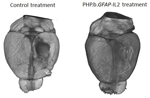
The new therapeutic designed by the research teams was able to boost the levels of the survival molecule IL2 in the brain, up to the same levels found in the blood. This allowed the number of regulatory T cells to build up in the brain, up to 10-fold higher than normal. To test the efficacy of the treatment in a mouse model that closely resembles traumatic brain injury accidents, mice were given carefully controlled brain impacts and then treated with the IL-2 gene delivery system. The scientists found that the treatment was effective at reducing the amount of brain damage following the injury, assessed by comparing both the loss of brain tissue and the ability of the mice to perform in cognitive tests.

Lead author, Dr Lidia Yshii, explained: “Seeing the brains of the mice after the first experiment was a ‘eureka moment’ – we could immediately see that the treatment reduced the size of the injury lesion”.

Recognising the wider potential of a drug capable of controlling brain inflammation, the researchers also tested the effectiveness of the approach in experimental mouse models of multiple sclerosis and stroke. In the model of multiple sclerosis, treating mice during the early symptoms prevented severe paralysis and allowed the mice to recover faster. In a model of stroke, mice treated with the IL2 gene delivery system after a primary stroke were partially protected from secondary strokes occurring two weeks later.  In a follow-up study, still undergoing peer review, the research team also demonstrated that the treatment was effective at preventing cognitive decline in ageing mice.

“By understanding and manipulating the immune response in the brain, we were able to develop a gene delivery system for IL-2 as a potential treatment for neuroinflammation. With tens of millions of people affected every year, and few treatment options, this has real potential to help people in need. We hope that this system will soon enter clinical trials, essential to test whether the treatment also works in patients." said Prof. Liston.

Dr Ed Needham, a neurocritical care Consultant at Addenbrooke’s Hospital who was not a part of the study, commented on the clinical relevance of these results: “There is an urgent clinical need to develop treatments which can prevent secondary injury that occurs after a traumatic brain injury. Importantly these treatments have to be safe for use in critically unwell patients who are at high risk of life-threatening infections. Current anti-inflammatory drugs act on the whole immune system, and may therefore increase patients' susceptibility to such infections. The exciting progress in this study is that, not only can the treatment successfully reduce the brain damage caused by inflammation, but it can do so without affecting the rest of the body's immune system, thereby preserving the natural defences needed to survive critical illness."

Thursday
May262022

Baanbrekende nieuwe behandeling leidt tot beter herstel na hersentrauma bij muizen

Onderzoekers hebben een gerichte therapie ontworpen die ontsteking in de hersenen tegengaat. De aanpak – waarbij doelgericht DNA tot in de hersenen wordt gebracht - blijkt succesvol bij muizen met een hersenletsel, beroerte of multiple sclerose

Kort & bondig:

  • Onderzoekers hebben een gerichte therapie ontworpen die ontsteking in de hersenen tegengaat. De aanpak – waarbij doelgericht DNA tot in de hersenen wordt gebracht - blijkt succesvol bij muizen met een hersenletsel, beroerte of multiple sclerose. 
  • De behandeling bestaat er in het aantal regulatoire T-cellen, die de anti-inflammatoire respons van het immuunsysteem reguleren, te verhogen in de hersenen.  
  • Door het aantal regulerende T-cellen in de hersenen te verhogen, konden de onderzoekers het afsterven van hersenweefsel bij muizen na verwonding voorkomen en deden de muizen het beter bij cognitieve testen. 
  • Voor patiënten met een traumatisch hersenletsel zijn er momenteel weinig opties om schadelijke neuroinflammatie te voorkomen. De nieuwe resultaten zijn dus erg hoopgevend. 

Onderzoekers uit Leuven en uit het Verenigd Koninkrijk publiceren vandaag nieuwe resultaten over een therapeutische piste waarbij het immuunsysteem wordt ingezet om hersenbeschadiging te voorkomen. De samenwerking tussen professor Adrian Liston (voorheen VIB en KU Leuven en sinds enkele jaren verbonden aan het Babraham Institute in het VK) en professor Matthew Holt (verbonden aan VIB, KU Leuven en de i3S-Universiteit van Porto) leidde tot een systeem om in de hersenen het aantal gespecialiseerde ontstekingsremmende immuuncellen op te voeren om op die manier hersenontsteking en -beschadiging te beperken. De aanpak bleek alvast in muismodellen tot minder hersenschade te leiden na hersenletsel, beroerte of bij multiple sclerose. Het onderzoek wordt vandaag gepubliceerd in het tijdschrift Nature Immunology. 

Traumatisch hersenletsel, zoals veroorzaakt na een auto-ongeval of een val, is wereldwijd een belangrijke doodsoorzaak. Het kan langdurige en ernstige gevolgen hebben voor mensen die het overleven, onder de vorm van cognitieve problemen en zelfs dementie. Een belangrijke oorzaak van deze cognitieve stoornissen is de ontstekingsreactie op het letsel. Hierbij veroorzaakt zwelling van de hersenen permanente schade. Terwijl ontsteking in andere delen van het lichaam kan worden aangepakt met geneesmiddelen, is dit in de hersenen heel moeilijk door de aanwezigheid van de bloed-hersenbarrière, die voorkomt dat gewone ontstekingsremmende moleculen op de plaats van het (hersen)trauma kunnen komen.  

Prof. Liston: "Ons lichaam heeft zijn eigen ontstekingsremmende respons: regulatoire T-cellen zijn in staat om ontstekingen waar te nemen en een cocktail van natuurlijke ontstekingsremmers te produceren. Helaas zijn er maar heel weinig van deze regulerende T-cellen in de hersenen. Wij probeerden een nieuw therapeutisch middel te ontwikkelen om de hoeveelheid regulerende T-cellen in de hersenen te vergroten. Indien er voldoende regulatoire T-cellen zijn, zo redeneerden we, zouden ze de ontsteking na een letsel kunnen beheersen en de schade beperken." 

Het onderzoeksteam ontdekte dat het aantal regulerende T-cellen in de hersenen laag was door een beperkte aanvoer van het cruciale overlevingsmolecuul interleukine 2, ook bekend als IL2. Het niveau van IL2 is laag in de hersenen vergeleken met de rest van het lichaam omdat het niet doorheen de bloed-hersenbarrière kan.  

Samen bedacht het team een nieuwe therapeutische aanpak waardoor meer IL2 kan worden aangemaakt door hersencellen. De onderzoekers gebruikten een "gene delivery"-systeem op basis van een virale vector: dit systeem kan daadwerkelijk een intacte bloed-hersenbarrière passeren en het DNA afleveren dat de hersenen nodig hebben om meer IL2 aan te maken.  

Prof. Holt: "Jarenlang leek de bloed-hersenbarrière een onoverkomelijke hindernis voor de efficiënte toediening van biologische geneesmiddelen in de hersenen. Ons werk, waarbij we gebruik maken van de nieuwste virale vectortechnologie, bewijst dat dit niet langer het geval is; het is zelfs mogelijk dat de bloed-hersenbarrière onder bepaalde omstandigheden therapeutisch gunstig kan zijn, omdat ze – eenmaal op hun bestemming – het 'lekken' van geneesmiddelen naar de rest van het lichaam verhindert." 

Dankzij hun aanpak waren de onderzoekers in staat de niveaus van de overlevingsmolecule IL2 in de hersenen op te voeren tot dezelfde niveaus als in het bloed. Hierdoor kon het aantal regulerende T-cellen zich in de hersenen opbouwen, tot 10 maal hoger dan normaal. Om de doeltreffendheid van de behandeling te testen in muizen. Wat bleek? Muizen met meer IL2 hadden inderdaad minder hersenschade na een letsel en presteerden ook beter in cognitieve tests.  

Dr. Lidia Yshii, van het team aan KU Leuven, legt uit: "Toen we de hersenen van de muizen zagen na het eerste experiment, was dit een echt 'eureka-moment' - we zagen meteen dat de behandeling het letsel had verkleind."  

De onderzoekers testten ook de doeltreffendheid van hun aanpak in experimentele muismodellen voor multiple sclerose en beroerte—met succes. In een vervolgstudie, die nog aan peer review wordt onderworpen en dus nog niet gepubliceerd is, toont het onderzoeksteam ook aan dat de behandeling doeltreffend was om cognitieve achteruitgang bij ouder wordende muizen te voorkomen.  

"Door de immuunrespons in de hersenen te begrijpen en erop in te spelen, waren we in staat een gen-toedieningssysteem voor IL-2 te ontwikkelen als een potentiële behandeling voor neuro-inflammatie. Met tientallen miljoenen mensen die er elk jaar mee te maken krijgen en met bovendien weinig beschikbare behandelingsmogelijkheden, biedt onze nieuwe aanpak reële mogelijkheden om mensen in nood te helpen. We hopen dat dit systeem binnenkort aan klinische proeven zal worden onderworpen, die essentieel zullen zijn om te testen of de behandeling ook bij patiënten werkt," aldus Prof. Liston. 

Dr. Ed Needham, een neuroloog in het Addenbrooke's Hospital in het VK die geen deel uitmaakte van de studie, gaf commentaar op de klinische relevantie van deze resultaten: "Er is een dringende klinische noodzaak om behandelingen te ontwikkelen die secundair letsel kunnen voorkomen dat optreedt na een traumatisch hersenletsel. Belangrijk is dat deze behandelingen veilig zijn voor gebruik bij kritisch zieke patiënten die een hoog risico lopen op levensbedreigende infecties. De huidige ontstekingsremmende geneesmiddelen werken in op het gehele immuunsysteem en kunnen daardoor de gevoeligheid van patiënten voor dergelijke infecties vergroten. Wat deze studie zo interessant maakt is dat de behandeling niet alleen met succes de door ontsteking veroorzaakte hersenschade kan verminderen, maar dat zij dit kan doen zonder de rest van het immuunsysteem van het lichaam aan te tasten, waardoor de natuurlijke afweer die nodig is om kritieke ziekte te overleven, behouden blijft." 

Thursday
Feb032022

Manipulating brain Tregs to protect against neuropathology

From the GlobalImmuno Talks 20222:

Tuesday
Jan122021

Top 10 health innovations of 2020

Great to see our recent Cell paper on brain T cells licensing microglia listed as one of the top 10 health innovations of 2020!

Thursday
Jul232020

Today in Cell


Not a bad day for the VIB Brain and Disease department...


Wednesday
Jul222020

New role for white blood cells in the developing brain

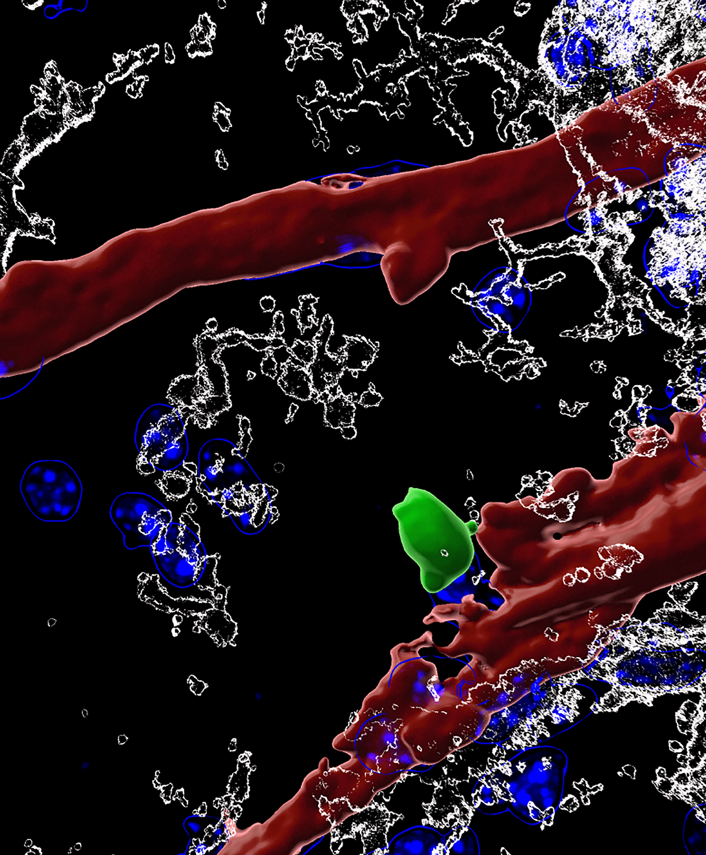
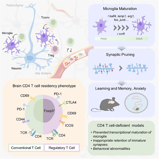
Whether white blood cells can be found in the brain has been controversial, and what they might be doing used to be complete mystery. In a seminal study published in Cell, an international team of scientists led by Prof. Adrian Liston (VIB-KU Leuven, Belgium & Babraham Institute, UK) describe a population of specialized brain-resident immune cells discovered in the mouse and human brain, and show that the presence of white blood cells is essential for normal brain development in mice.

Like a highly fortified headquarters, our brain enjoys special protection from what is circulating in the rest of our body through the blood-brain barrier. This highly selective border makes sure that passage from the blood to the brain is tightly regulated.

The blood-brain barrier also separates the brain from our body’s immune system, which is why it has its own resident immune cells, called microglia, which trigger inflammation and tissue repair. Microglia arrive in the brain during embryonic development, and later on, the population becomes self-renewing.

Yet, white blood cells—which are part of our immune system—have been found to play a role in different brain diseases, including multiple sclerosis, Alzheimer’s and Parkinson’s disease or stroke. Whether or not white blood cells can be found in healthy brains as well, and what they might be doing there, has been subject of intense debate. An interdisciplinary team of scientists led by Prof. Adrian Liston (VIB-KU Leuven, Babraham Institute) set out to find the answers.

White blood cells in the brain

"A misconception about white blood cells comes from their name,” explains Dr. Oliver Burton (Babraham Institute). “These 'immune cells' are not just present in the blood. They are constantly circulating around our body and enter all of our organs, including—as it turns out—the brain. We are only just starting to discover what white blood cells do when they leave the blood. This research indicates that they act as a go-between, transferring information from the rest of the body to the brain environment"

The team quantified and characterized a small but distinct population of brain-resident T helper cells present in mouse and human brain tissue. T cells are a specific type of white blood cells specialized for scanning cell surfaces for evidence of infection and triggering an appropriate immune response. New technologies allowed the researchers to study the cells in great detail, including the processes by which circulating T cells entered the brain and began to develop the features of brain-resident T cells.

Dr. Carlos Roca (Babraham Institute): “Science is becoming increasingly multidisciplinary. Here, we didn't just bring in expertise from immunology, neuroscience and microbiology, but also from computer science and applied mathematics. New approaches for data analysis allow us to reach a much deeper level of understanding of the biology of the white blood cells we found in the brain.”

An evolutionary role

When T helper cells are absent from the brain, the scientists found that the resident immune cells – microglia – in the mouse brain remained suspended between a fetal and adult developmental state. Observationally, mice lacking brain T cells showed multiple changes in their behavior. The analysis points to an important role for brain-resident T cells in brain development. If T cells participate in normal brain development in mice, could the same be true in humans?

“In mice, the wave of entry of immune cells at birth triggers a switch in brain development,” says Liston. “Humans have a much longer gestation than mice though, and we don't know about the timing of immune cell entry into the brain. Does this occur before birth? Is it delayed until after birth? Did a change in timing of entry contribute to the evolution of enhanced cognitive capacity in humans?”

The findings open up a whole new range of questions about how the brain and our immune system interact. "It has been really exciting to work on this project. We are learning so much about how our immune system can alter our brain, and how our brain modifies our immune system. The two are far more interconnected than we previously thought," says Dr. Emanuela Pasciuto (VIB-KU Leuven).

The study also brings in a connection with the gut microbiome, says Liston: “There are now multiple links between the bacteria in our gut and different neurological conditions, but without any convincing explanations for what connects them. We show that white blood cells are modified by gut bacteria, and then take that information with them into the brain. This could be the route by which our gut microbiome influences the brain.” 

Taken together, the results contribute towards the increasing recognition of the role of immune cells in the brain and shed new light on its involvement in a range of neurological diseases.

Check out the full article here

Wednesday
Jul222020

Witte bloedcellen ook belangrijk voor ons brein

Witte bloedcellen maken deel uit van ons immuunsysteem dat ons tegen ziektes beschermt.  Of ze ook in de hersenen terug te vinden zijn, en wat ze daar dan zouden doen bleef tot nog toe een raadsel. Een internationaal team van wetenschappers onder leiding van professor Adrian Liston (VIB-KU Leuven, België en Babraham Institute, VK) toont nu aan dat witte bloedcellen wel degelijk voorkomen in de hersenen van zowel muizen als mensen, en dat hun aanwezigheid essentieel is voor normale hersenontwikkeling. De resultaten verschijnen deze week in het prestigieuze vakblad Cell.

Onze hersenen worden als een versterkte burcht ommuurd door de bloed-hersenbarrière. Die moet vermijden dat stoffen die in onze bloedbaan circuleren zomaar in onze hersenen terechtkomen. Via de bloed-hersenbarrière worden onder strikte controle enkel welbepaalde stoffen uitgewisseld.

De bloed-hersenbarrière schermt de hersenen ook af van ons immuunsysteem, dat de rest van ons lichaam patrouilleert om bijvoorbeeld bacteriële of virale indringers op te sporen en uit te schakelen. Precies daarom heeft het brein z’n eigen immuuncellen: microglia.

Toch blijken witte bloedcellen ook een rol te spelen bij verschillende hersenaandoeningen. Denk maar aan MS, alzheimer, parkinson of een beroerte. Hierbij gaat het wel telkens om ziek of ‘beschadigd’ hersenweefsel, waar mogelijk ook de bloed-hersenbarrière is aangetast. De vraag bleef dus of – en waarom – witte bloedcellen nu werkelijk aanwezig zijn in hersenen die normaal en gezond zijn.

Witte bloedcellen in de hersenen

Een interdisciplinair team van wetenschappers onder leiding van prof. Adrian Liston (VIB-KU Leuven, Babraham Institute) heeft nu een kleine maar belangrijke groep van T-helpercellen ontdekt in hersenweefsel afkomstig van muizen en van mensen. T-helpercellen zijn een specifiek type witte bloedcellen, gespecialiseerd in het scannen van celoppervlakken op aanwijzingen van infectie en in het op gang trekken van een aangepaste immuunreactie. Aan de hand van de laatste technologie konden de wetenschappers de T-helpercellen tot in detail bestuderen, inclusief hoe en wanneer ze in de hersenen terecht komen.

Dr. Emanuela Pasciuto (VIB-KU Leuven), postdoctoraal onderzoeker in het team van Liston benadrukt het belang van interdisciplinair onderzoek: “Om de rol van witte bloedcellen in het brein in kaart te brengen hebben we niet alleen expertise van immunologie, neurowetenschappen en microbiologie bij elkaar gebracht, maar ook van informatica en toegepaste wiskunde. Nieuwe benaderingen voor data-analyse stellen ons in staat om een ​​veel dieper begrip te krijgen van de biologie van de witte bloedcellen die we in de hersenen hebben gevonden.”

Een evolutionaire rol

De onderzoekers stelden vast dat in muizenhersenen zonder T-helpercellen de ontwikkeling van de typische immuuncellen van het brein (de microglia) bleef hangen ergens tussen een foetale en volwassen ontwikkelingsstatus. De muizen zonder T-helpercellen in de hersenen vertoonden bovendien verschillende gedragsafwijkingen, wat wijst op een belangrijke rol voor de T-helpercellen bij de normale hersenontwikkeling. En als dat geldt voor muizen, zou hetzelfde dan ook waar zijn voor mensen?

“We zien dat de toestroom van immuuncellen in de hersenen bij de geboorte van muizen leidt tot een omslag in het ontwikkelingsproces,” zegt Liston. “Maar de zwangerschap bij mensen is veel langer dan bij muizen, en we weten niet wanneer de immuuncellen dan toekomen in het menselijk brein. Gebeurt het nog vóór de geboorte? Is het uitgesteld tot na de geboorte? Kan een verandering in de timing bijgedragen hebben aan de evolutie van de uitzonderlijke hersencapaciteit van mensen?”

De bevindingen openen een heel nieuw gamma aan vragen over de wisselwerking tussen ons brein en ons immuunsysteem. “We leren nog elke dag bij over hoe ons immuunsysteem ons brein kan beïnvloeden en vice versa. De twee zijn veel meer met elkaar verbonden dan we eerder dachten,” zegt Pasciuto.

Darmen en hersenen

De studie legt ook nieuwe verbanden tussen ons brein en onze darmflora, aldus Liston: “Heel wat neurologische aandoeningen worden in verband gebracht met bacteriën in onze darmen, maar zonder overtuigende verklaringen voor die connectie. Onze resultaten laten zien dat darmbacteriën witte bloedcellen kunnen beïnvloeden, die deze ‘informatie’ vervolgens mee nemen naar de hersenen. Dit zou de manier kunnen zijn waarop onze darmflora onze hersenen beïnvloeden. ”

De nieuwe resultaten dragen enorm bij tot de groeiende kennis over de rol van immuuncellen in de hersenen, zowel tijdens de normale ontwikkeling als bij verschillende ziekteprocessen.